|
Thanatology
,the science dealing with the study of death has been inexistence for
centuries. It is interesting to review some of the notions that
prevailed earlier. Legal and medical quandaries regarding the
definition of death are not new. In his Historia Naturalis, the Roman
author Pliny the Elder wrote that "so uncertain" is men’s
judgment that they cannot determine even death itself.
Perceptions of death have been reflected in poetry,
literature, legends and pictorial art. Human beings have been the
only species to bury their dead in a systematic way, often with
implements to be used in a further existence. Most ancient
civilizations (Egyptian, Zoroastrian, Hindu, Christian, Judaic and
Islamic) however accepted death as an easily determined empirical
fact, not requiring or further elaboration.
Today a conceptual crisis has arisen in modern medicine
and biology. This crisis stems precisely from the realization that
the definition if death – taken for granted by our ancestors requires
re examination. Death from a biological angle necessarily has to be
redefined. Many dictionaries define death as "the extinction or
cessation" if life or as ceasing to be. Today death of the brain
is considered to be the death of the individual and death of the
brain stem, is accepted as death of the brain and therefore of the
individual.
Death A Process or an Event ?
Is death the irreversible loss of function of the whole
organism, that is, of everyone of its component parts? Or is it the
irreversible loss of function of the organism as a whole; that is
loss of the ability to exist as a meaningful and independent
biological unit? Civilizations fall apart yet their component societies
live on; societies disintegrate but their citizens survive;
individuals die while their cells, perversely, still metabolize;
finally, cells can be disrupted yet the enzymes they release may, for
a while, remain very active. So what is death and when is a person
considered dead?
One must realize and accept that death is a continuous
ongoing process, not an isolated event. To certify that death
occurred at a specific time is not, from a purely biological and
cellular point of view, acceptable. Until recently however this had
no practical implications. Death of different organs x(-) the heart,
the brain and so on occurred rapidly within seconds of each other. A
few decades ago technology was not available to keep some organs
alive even though others were dead. Unless caught up in nuclear
explosions people do not die instantaneously, like the bursting of a
bubble. Several minutes after the heart has stopped beating,
electrical activity can still be recorded if one probes for signals
from within the cardiac activity. Three hours after death the pupils
still respond for signals from within the cardiac activity. Three
hours after death the pupils still respond to pilocarpine drops by
contracting, and muscles repeatedly tapped may still mechanically
shorten. Kidneys can be removed even two hours after irreversible
cardiac arrest. Bones taken 48 hours after death can still be
transplanted. Arteries can be grafted as late as 72 hours after the
heart has irreversibly stopped cells clearly differ widely in their ability
to withstand the deprivation of oxygen supply that follows arrest of
the circulation. The challenge is identical such points with greater
precision for different organs.
At the clinical level the irreversible cessation of
circulation has for centuries been considered the point of no return.
It has provided (and still provides) a practical and valid criterion
of irreversible loss of function of the organism as a whole. What is
new is a awareness that circulatory arrest is a mechanism of death; that
cessation of the heart beat is only lethal if it lasts long enough to
cause critical centers in the brain stem to die; and that this is so
because the brain stem is irreversible in a way the cardiac pump is
not. These are not so much new facts as new ways of looking. At old
ones.
Cell Death:
Programmed cell death plays an important role in
embryological development and teratogenesis. Such programmed events
are essential if the organism as a whole is to develop to its normal
final form. Waves of genetically driven cell death are critical to
the proper modeling of organs and systems. The infections of the
developing mammalian brain and spinal cord is due to death of cells
at appropriate times. Programmed cell death may also play a part in
the process of aging cells which are designed to die after a certain
number of cell divisions.
Human death cannot be simplified to purely biological
terms, divorced from ethical and cultural considerations. The
repercussions (burial mourning, inheritance, etc.,) are many. They
have to be socially acceptable in a way that does not apply to the
fate of cells in tissue culture. Technical data can never answer
purely conceptual questions. Capacity for consciousness is a function
of the brainstem while content of consciousness is a function for the
cerebral hemispheres. If there is no functioning brain stem there can
be no meaningful or integrated activity of the cerebral hemispheres,
no social interaction with the environment, nothing that might
legitimize ending the adjective sapiens (wise) ti the noun Homo
(man). The capacity for consciousness is per haps the nearest one can
get to giving a biological flavor to the notion of the soul.
Pope Pius XII, speaking to an International Congress of
Anesthesiologists in 1957, raised the question of when, in the
intensive care unit, the soul actually left the body. More secularly
inclined philosophers have meanwhile pondered what it was that was so
essential to the nature of man that its loss should be called death.
English author Sir Thomas Browne in 1643 remarked : With what strife
and pains we cine come into the world we know not, but it is commonly
no easy matter to get out of it.
History of Brain Death:
Brain death was first described by two French
physicians, Mollart and Goulon and termed coma depasse (a state
beyond coma) They differentiated coma depasse from coma prolonged,
the latter being the condition, which is now termed persistent
vegetative state. In 1968 the Ad Hoc Committee of the Harvard Medical
School defined brain death as irreversible coma, with the patient
being totally unreceptive and unresponsive, with absent reflexes and
no spontaneous respiratory effort during a 3 min period of
disconnection from the ventilator. The report unambiguously proposed
that this clinical state should be accepted as death. A few years
later Mohandas and Chou suggested that in patients with know but
irreparable intracranial lesions, reversible damage to the brainstem
was the point of no return and that the diagnosis could be based on
clinical judgment, thereby introducing the important concepts of
etiological preconditions and a purely clinical diagnosis. Another
important contribution was the memorandum issued by the Conference of
Royal Medical Colleges (1976). This emphasized that permanent functional
death of the brainstem constitutes brain death and that this should
only be diagnosed in the context of irremediable structural brain
damage, after exclusion of certain specified conditions, which might
contribute to or cause the coma.
A second memorandum issued in 1979 equated brainstem
death with death itself. Therefore death can be declared once death
of the brainstem has been confirmed, and most would argue that
mechanical ventilations should then be discontinued as soon as
possible. This should be be viewed as withdrawing support to allow a
patient to die, but rather as ceasing a futile intervention in a
patient who is already dead. Therefore it is clear that, even if
transplantation therapy did not exist, the ability to diagnose brain
death with confidence contributes to the humane practice of intensive
care, and most clinicians find the decision to discontinue
ventilating a brain dead patient relatively straightforward. What was
clearly established in the early 1980s was that no patient in apnoeic
coma declared brain dead, according to the very stringent criteria of
the UK code (outlined in the 1976 and 1979 Memoranda of the
conference of Royal Medical Colleges) had ever regained consciousness
or had ever failed to develop asystole within a relatively short
time. The acceptance of these ideas would lessen human distress, lead
to more rational use of limited intensive are facilities, and
radically after the life expectancy of thousands of patients with end
stage organ failure waiting desperately for organs.
How much of the brain needs to be destroyed to produce
death? The destruction of a crucial few cubic centimeters of tissue
lying beneath the aqueduct of Sylvius anteriorly and in the floor of
the fourth ventricle posteriorly, is all that is required to ensure
irreversible loss of brain function. The concept of brainstem death
became operational in India after the enactment of legislation by the
Indian Parliament and its notification in the Gazette to India. It
recognizes brainstem death based on the UK criteria, which have the
advantage of being simple, clinical, unequivocal and capable of
confirmation.
Brain death should not even be thought of until the
following reversible causes of coma have been excluded :
Intoxication (alcohol)
Drugs, which depress the central nervous system.
Muscle relaxants
Primary hypothermia (by measuring rectal temperature)
Hypovolaemic shock (by sequential measurement of blood
pressure)
Metabolic and endocrine disorders.
Incidence of brain death:
Walker is quoted as having stated that brain death
occurs in approximately 1% of all deaths. According to Jennett et al,
the occurrence, with about 4000 cases occurring each year in Britain.
Pathophysiology of brain death:
The changes in the brain following brain death are a function
of time. The pathogenesis includes direct cellular injury potentiated
by a vicious cycle of failure of blood flow, hypoxia, cerebral
acidosis and endothelial swelling to brain edema, herniation and
aseptic necrosis of the brain. Gross examination of such brain
specimens shows a dusky, congested cerebral cortex, generalized brain
swelling, a swollen pituitary gland and macerated cerebellum.
Microscopically, there is pan-necrosis of the nervous tissue and
extensive foci of necrosis throughout the cerebrum brainstem and
cerebellum, The physiological changes following brain death are so
severe the progressive somatic deterioration and cardiac standstill
will inevitably occur despite extensive life support. A number of
subsequent studies have suggested that brain death does not always
rapidly lead to somatic death. In one series, cardiac rhythm could be
maintained for prolonged periods (mean (SD) duration < or=23.1
(19.1) days) after the declaration of brain death.
Physiological changes:
The physiological changes occurring in organs distant
from the brain at or around the time of onset of brain death arise as
a result of two major mechanisms.
Diffuse injury to the vascular regulation mechanism
occurring due to early massive sympathetic outflow, followed by its
profound reduction.
Diffuse metabolic cellular injury due to lack of
hypothalamic control, producing generalized metabolic and hypoxic
lesions in all tissues.
Circadian changes in temperation (high at day, low at
night) however are preserved during the period of brain death.
Reduced intraocular pressure is a feature of brain death
(12/12). Pallor or disc suggestive of funds ischemia was common. Disc
edema was not noted.
Physiological, histological , biochemical and ECG
evidence of damage to the heart has been documented at the time of
brain death. The sudden increase in ICP and resultant cerebral
ischemia leads to an autonomic or sympathetic storm due to massive
outpouring of catecholamines (Cushing’s reflex). The rise in
catecholamines depends on the rate of rise in ICP. There is an
initial increase in parasympathetic tone with bradycardia, followed
by marked sympathetic changes leading to hypertension, tachycardia a
vasoconstriction. Immediately after the autonomic storm, there is
loss of cardiovascular tone with brady arrythmias, vasodilation and
consequent hypotension, Hypotension and low caridac output then start
a cycle of poor myocardial and tissue perfusion with further decrease
in myocardial performance. In a few centers, brain dead organ donors
are not considered for heart donation if high levels of adrenaline
are required to maintain cardiovascular variables within normal
limits.
Neuropathology of the persistent vegetative state as
distinct from neuropathology in brain death has been reviewed.
Hypernatremia, diabetes Insipidus is more often the
effect rather than the cause.
Maintenance of the brain dead mother to ensure viability
of the fetus is fraught with major problems and is extremely
expensive but can be done.
Clinical evaluation of brain stem death :
In more and more countries, certification of brain stem
death is made on purely clinical grounds. The aim of the clinical
test is not to probe every neuron within the intracranial cavity to
see if it is dead" an impossible task but to establish
irreversible loss of brain – stem function. The accuracy,
reliability, reproducibility and ease in carrying out clinical tests
make clinical evaluation sufficient for diagnosis of brainstem death.
The President’s Commission also outlined guidelines for the
determinations of death for the Study of Ethical Problems in Medicine
and Behavioral Research. Neuro physiological and imaging studies are
not essential to confirm brain death. By testing various brain stem
reflexes, the functions of the brain stem can be assessed clinically
with an ease, thoroughness, and degree of detail not possible for any
other part of the central nervous system.
Pupillary response to light: The
response to bright light should be absent in both eyes. The pupil
should be observed closely for one minute to allow time for a slow
response to become evident. Both widely dilated as well as
mid-positioned fixed pupils are seen in brain dead patients. The size
may vary from 4 – 9 mm. Widely dilated pupils are not a necessary
criterion for brain death but fixed pupils with no response to light
are mandatory.
Corneal reflex: This should be absent.
Repeated corneal stimulation is unnecessary and should be avoided
=Corneal abrasions are undesirable if the patient is a potential
corneal donor.
Fifth and Seventh Cranial Nerves: There
should be no motor response in the distribution of any cranial nerve.
Such a response would be grimacing (facial nerve motor response) in
response to thumb pressure over the supra orbital groove (trigeminal
nerve sensation). Similarly, there should be no response to painful
stimuli of the trunk suggesting absence of sensory nerve conduction
across the foramen magnum.
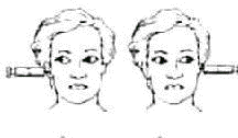
Oculo cephalic reflex (Doll’s eye phenomenon): This
test must not be performed in patients with an unstable cervical
spine. The head is turned from starting position to a new steady
position and briskly to the opposite side. The eyes move as shown
in…. denoting the integrity of the medial longitudinal fasciculus in
the brain stem .
Gag reflexes: This should be absent. A
tongue depressor is used to stimulate each side of the oropharynx and
the patient observed for any pharyngeal or palatal movement.
Evaluation of Gag reflex may be difficult in an intubated patient and
should not be performed if extubation is required.
Cough reflex: A suction catheter is
introduced into the endotracheal or tracheostomy tube to deliberately
stimulate the carina. The patient is closely observed for any cough
response or movement of the chest or diaphragm.
Oculovestibular reflex: Before
testing, both ears must be inspected with an auroscope to confirm
that the tympanic membranes are intact and the external auditory
canal not obstructed. If the eardrum is perforated, the test can be
performed using cold air as the stimulus. A fracture of the base of
skull resulting in blood, cerebrospinal fluid or brain tissue in the
external auditory canal is a contraindication to performing this test
on that ear. The patient’s head is placed in the center and lifted 30
degree from the supine position. A soft catheter is introduced into
the external auditory canal and slow irrigation with at least 5-ml of
ice-cold water is performed while, the eyes are held open by an
assistant. The eyes should be observed for one minute after
irrigation is completed before repeating the test on the other side.
An intact oculovestibular reflex causes tonic deviation of the eyes
towards the irrigated ear. Any movement of one or both eyes, whether
conjugate or not, excludes the diagnosis of brain death. In a brain
dead patient the eyes remain fixed. Combined ice-cold water caloric
stimulation and head rotation has been suggested as the most
pro-found stimulation for deeply unconscious patients.
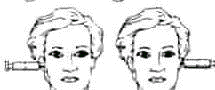
Apnoea test: Apnoea testing is
essential for confirmation of brain death. It should only be done
when all the prerequisites have been met and all other brain stem
reflexes are absent. It is not possible to perform this test in a
patient with high cervical cord injury, which may have abolished
phrenic nerve function. Important changes in vital sings (ex; marked
hypotension, servere cardiac arrhythmias) during the apnoea test may
be related to lack of adequate precautions, although they may occur
spontaneously during increasing acidosis.
Therefore, the following prerequisites have been
suggested.
The core temperature should be> or
= 36.5 degree Celsius; The systolic blood pressure should be
> or = 90mm Hg; Euvolaemia (preferably positive fluid
balance in the previous (6hour); Eucapnoea (arterial
pCO2>or=40mmHg). A useful method of raising the pCO2 in an over
ventilated hypocapnic patient is to connect an oxygen filled bag to
the endotracheal tube and rebreathe pure oxygen for 10 minutes
without CO2 exhaustion.
The three components of the apnoea test are:
Absence of spontaneous respiratory efforts during a
period of disconnection (10 min.) from the mechanical ventilator.
Arterial carbon dioxide must reach a critical
point(>60mmHg) during this period.
Prevention of hypoxemia during this period.
The steps in testing are :
Disconnect the ventilator
Deliver 100% oxygen at 6 L/min; place a cannula at the
level of the carnia.
Look closely for respiratory movements. Respiration is
defined as abdominal or chest excursions that produce adequate tidal
volumes. Respiratory – like movements can occur at the end of the
apnoea test, when oxygenation may become marginal. However, these do
not produce adequate tidal volumes. When the test is in doubt, a
spirometer can be connected to the patient to confirm the absence of
tidal volumes.
Measure arterial pO2, pCO2, and pH after 10 minutes and
reconnect the ventilator.
If the respiratory movements are absent and the arterial
pCO2>or 60 mmHg (20mmHg increase in pCO2 over baseline) the apnoea
test is positive), i.e. it supports the diagnosis of brain death.
If respiratory movements are observed, the apnoea test
is negative (i.e. it does nto support the clinical diagnosis of brain
death), and the test should be repeated.
If during the apnoea test the systolic blood pressure
becomes < or = 90 mmHg, the pulse oximeter indicates marked
desaturation, and cardiac arrhythmias occur, draw a blood sample
immediately, connect the ventilator and analyze arterial blood gases.
The apnoea test is positive if the arterial pC)2 us > or=60mmHg.
If the pCO2 is <60mmHg, the resul;t is indeterminate and repeat
testing at a later stage should be done.
|
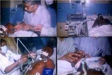
Tests to confirm brain death:
The plethora of gadgetry ultimately only gives answers
of dubious reliability to the wrong questions! None are superior
to clinical assessment At present, there is no evidence that, MRI,
MRA, EEG, evoked potentials, Trans Cranial Doppler, evaluation of
cerebral blood flow or other tests can unequivocally
establish brainstem death. These techniques though under review,
currently do not form part of the mandatory diagnostic requirements
in most countries. Some countries however include these tests.
|
|
|
|
|
|
|
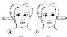
Retained
Vestibulo-ocular reflexes
|
|
|
Rt
(a) & Lt (b) 6th nerve palsies
|
|
|
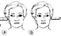
Lt(a)
& Rt (b) internuclear ophthamoplegia
|
|
|
Absent
Vestibulo-ocular reflexes
|
|
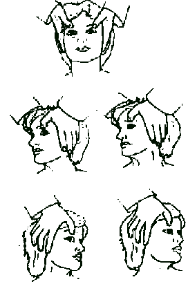
Testing for Doll's eye movements
|
EEG and Evoked Potentials:
Some still consider that, demonstration of absence of
cerebral electrical activity, is necessary to diagnose brain death.
Others recommend the use of evoked potentials to assist in the
diagnosis of brain death since these can be demonstrated when EEG
silence is attributable to drugs. Evoked potential was preserved in
coma in all patients, but lost in brain death in 100%. It is
therefore useful in distinguishing isolated brainstem death from high
cervical transverse cord lesions and focal bilateral lemniscal
lesions. In the UK most argue that the surface EEG cannot exclude
activity in deeper areas of the brain, EEG may also not show
electrical activity in barbiturate coma. Patients have been reported
in whom the EEG was isoelectric but brainstem reflexes were
preserved, although this is extremely unusual.
MRI and brain death
Fifteen patients with clinical diagnosis of brain death
were examined by MRI. MRI showed that flow voids were absent in the
ICA in all eight patients in whom non-filling was confirmed by IADSA.
Partial residual flow voids may be caused by to and fro blood movement
which was demonstrated by transcranial Doppler sonography. Several
authors have commented on the role of MRI in the evaluation of brain
death. There are even reports on contrast enhanced CT changes in
brain death.
Cessation of Cerebral Blood Flow:
Clinical and electrophysiological criteria may be
misinterpreted due to drug intoxication, hypothermia or technical
artifacts. Thus, if clinical assessment is sub optimal, reliable
early confirmatory tests may be required for demonstrating absence of
intracranial blood flow. All patients with isolated brain lesions and
Glasgow Coma Scale (GCS) = 3 were subjected to neurological
examination after ruling out hypothermia, metabolic disorders and
drug intoxications and diagnosed as clinically brain dead when the
brainstem reflexes were absent and the apnoea test positive. Cerebral
blood flow measurements with the i.v. Xe-133 method (CBF) and
selective cerebral angiography were carried out. EEG was isoelectic
in 8 petients while the remaings 7 patients showed persistence of
electrical activity. Trans cranial Doppler was compatible with
intracranial circulatory arrest in 18 MCA districts, compatible with
normal flow in 2 and undetectable in 10 out of 30 districts
insonated. Cerebral Angiography and CBF studies are the most reliable
investigations whereas the role of EEG and TCD remain to be
determined because of the presence of false negatives and positives.
Cerebral blood flow velocities in the middle cerebral arteries were
measured using transcranial Doppler in 12 patients who had conditions
that ultimately resulted in brain death. This pattern consisted of
reverberating flow, with forward flow in systole and retrograde flow
in diastole. When this pattern was seen, there was arrest of cerebral
flow, as measured by radionuclide scanning Radionuclide cerebral
scanning cannot document absence of flow in the vertebrobasilar
circulation. Color flow duplex scanning may be used to complement
radionuclide cerebral scanning. Reports claiming superiority of
perfusion studies with Tc-99mHMPAO over conventional radionuclide
cerebral Angiography have been reported.
Transcranial Doppler was conducted transtemporally on
the left and right-middle cerebral artery four times daily. In all
patients, transcranial Doppler waveforms exhibited high resistance
profiles with low, zero, and then reversed diastolic flow
velocity-only three waveform patterns, consisting of diastolic
forward flow, brief systolic forward flow. This noninvasive method to
document deterioration of cerebral profusion pressure could be
included in the future in protocols for brain death diagnosis.
Drugs as a confounding factor in evaluation of brain
death:
Effects of drugs must be excluded before considering
brain death. Most centrally acting drugs depress respiration and
would be expected to affect apnoea testing of brain stem function.
The entry of drugs into the brain is also altered in some disease
states. However the effects of central depressants when there is
damage to the blood-brain barrier or brain is not clearly known..
Drug screens can assist in determining whether drugs are present, but
correct interpretation of the results depends on close liaison
between the clinical and laboratory staff. Life support systems must
be continued when a centrally active drug is present. Post traumatic
brain death may occur in patients treated with barbiturate for
elevated ICP. Saits et al report two cases of brain death where a
large amount of barbiturate remained in the brain, even when the
blood concentration was not detectable, possibly because the blood
flow was stagnant in the brain. It is suggested that a patient in
barbiturate coma should not be diagnosed to be brain dead.
Clinical observations compatible with brain death:
Spinal reflexes the spinal cord may continue to function
after the death of the brainstem. The resulting limb movement may
cause distress to both family and staff caring for the patient. After
the second set of brainstem death tests are completed and the patient
has been certified brain dead, muscle relaxants may be given for
spinal reflexes to prevent further distress to the family. Muscle
stretch, superficial abdominal and the Babinski reflexes are of
spinal origin and do not invalidate the diagnosis of brain
death.
Hemodynamic responses:
Profuse sweating, blushing, tachycardic and sudden
increases in blood pressure can be elicited by neck flexion in brain
dead patients.
Diagnosis of brainstem death:
Brainstem function evaluation should be performed
independently by at least two consultants either the consultant in
charge of the patient and another clinically independent of the first
and registered for more than 5 years. Neither should be a member of
the transplant team. Care, diligence and meticulous implementation of
established criteria are absolutely necessary. Criteria for
diagnosing Brain Death in Infants and Children (age of 2 months
should be the same as those in adults. The legal time of declaration
of brain death is the time at which brain death tests have been
repeated for a second time and found to be unequivocally positive.
The declaration of brain death must be recorded in the medical notes
with the date and time. In India the certification of death must
meningiomas as per the Transplantation of Human Organs Act.
Effect of brain death on the family:
Brain death has created a new class of dead people that
does not conform to society’s expectations of normal death and dying.
Brain death also causes great stress for the family and friends.
Effective communication, caring and supporting the family is crucial.
While these families have a variety of special needs, it is constant
interaction that provides opportunities to have a positive influence
on family member’s ability to cope with the tragedy and begin the
healing process.
Natural History of brain death:
The diagnostic mix of 1228 brain dead renal donors in
Britain was similar to that of 479 cases of brain death recently
reported form three neurosurgical units. About half the donors came
from non – teaching hospitals without a neurosurgical unit, many of
them small and distant from the center. Head injuries accounted for
half the donors, and intracranial hemorrhage for almost a third. This
suggests reluctance of doctors to initiate donation rather than
relatives withholding permission.
In three neurosurgical units, 609 patients diagnosed
clinically as brain dead were studied; 326 had final cardiac systole
while still being ventilated, and ventilation was discontinued in the
reminder. No patient recovered. The median time in hospital before the
heart finally stopped was 3-4 days, with 30-40 hours on the
ventilator. Analysis of prospective data from three countries on
patients with severe head injuries showed that not one of 1003
survivors would ever have been suspected of being brain dead even in
their worst state soon after injury.
MANAGEMENT OF THE POTENTIAL ORGAN DONOR:
Consequences of brain death:
Hypotension, Hypothermia
Requirement for multiple transfusions
Arrhythmias, Cardiovascular collapse
Cardiac arrest requiring CPR
Disseminated intravascular coagulation
Hypoxia, Pulmonary edema
Electrolyte Imbalances, Acidosis
Bacteremia, Infections
Diabetes Insipidus, Endocrine Disturbances,
Hyperglycaemia.
Assiduous supportive treatment of the brain dead
individual with due attention to the above, is essential to prevent
deterioration of initially suitable donors. Complications increase
progressively after brain death and, although adequate time must be
allowed to confirm the diagnosis, unnecessary delays must be avoided.
This will increase donation rates and improve graft
survival and function.
In organ retrieval aggressive supportive treatment of
the donor should be continued intra – operatively. Maintaining the
functioning of the salvageable body organs in a brain dead individual
is time consuming, complex, challenging and expensive.
A dead brain in a body with a beating heart is one of
the more macabre products of modern technology. During the past 30
years increasing use of more and more sophisticated ventilators,
pressor amines, intravenous alimentation and dialysis has resulted in
keeping alive organs, in a body whose brain has irreversibly ceased
to function, With increasing availability of intensive c are
facilities in India the number of brain deaths are likely to
increase. It is essential that the neurologist and neurosurgeon
recognize and confirm brain death. Once this has been done they
should not shy away, from discussing with the family the futility of
continuing support systems.
Possible organ donation should also be broached.
|
