|
Empty
sella is a radiological diagnosis based on CT or MR investigation.
Either a normal sized (empty sella) or enlarge sella (empty enlarged
sella) presents partly or totally filled with cerebrospinal fluid.
The radiological diagnosis does not mean a pathological situation in
every instance. Many patients present without specific symptoms and
the diagnosis is made by chance. Empty sella syndrome is the
pathological variant of a radio logically verified empty sella.
Primary empty sella is an
idiopathic form of an empty sella which occurs in the absence of prior
pituitary surgery or radiation therapy or medication with DOPA
agonists.
Secondary empty sella occurs as a
result of surgical resection or irradiation of a sellar expansion.
Anatomy:
The
disphragma sellae normally forms a circular fold that constitutes a roof
for the sella turcica with only a small central opening for the passage
of the pituitary stalk. Busch in 1951 performed an autopsy
study of 788 subjects without known pituitary disease. In 38.4% he
found a complete covering of pituitary gland by the diaphragm. He
observed an empty sella in 5.5%. Other studies described
significant defects in the sellar diaphragm in 22% to 72% of cases.
These defects were frequently accompanied by intrasellar extension of
subarachnoid space and downward displacement of the optic chiasm.
Busch
and others have measured the volume of the sella and of the sellar
contents. These measurements are widely variable because the definition
of the lateral and superior boundaries of the sella is arbitrary.
However, Bjerre(1990) concludes that a sella with a volume
exceeding 1.1-1.2cm3 calculated from X-ray is an enlarged
sella.
Incidence:
A
5:1 female male predominance exists in the incidence of diaphragmatic
defects.
Over
80% of the cases occur in women, the majority between the ages of 40 and
49. 78% to 90% of these patients were described as obese,
multiparous and 31% were hypertensive (Neelon, 1973).
An
empty sella of normal size without clinical significance was found in
some 10% (Brisman, 1978). In children radiological incidence of
primary empty sella was reported as 1-48% with a male-female ratio of
1.4:1.0 (Rappaport, 1991).
Etiology:
Primary empty sella
An
empty sella (either normal sized or enlarged) may simply be a normal
anatomical variation so that concurrent symptoms are unrelated to the
radiological appearance.
If
a deficient diaphragma sellae is present, increased intracranial
pressure, as in pseudotumor cerebri, displaces the suprasellar
arachnoidal and cerebral structures into the sellar cavity. This
may cause sellar enlargement and partial emptiness, as well as
posteroinferior displacement and compression of the pituitary gland, and
displacement of the optic chiasm and erosion of the sellar floor and
dura. The theory is supported by the following observations.
- An empty sella syndrome is observed frequently in
patients with benign intracranial hypertension. Weisberg
(1975) reported an incidence of 10% in 50 cases with benign
intracranial hypertension.
- Continuous intracranial pressure monitoring in
patients with empty sella syndrome revealed intermittent
asymptomatic increases in pressure in some patients (Kaye,
1982).
- Intracranial tumors of slow growth have been
associated with a deficient sellar diaphragma. The incidence
of an abnormal sellar configuration in patients with intracranial
tumors and empty sella syndrome is about 24%.
- Patients with known empty sella followed for some
years exhibit progressive changes of their radiological sellar
appearance.
Another
theory relates the empty enlarged sella to a previous pituitary
hyperplasia or hypertrophy (e.g. during pregnancy or because of primary
peripheral endocrine gland insufficiency). This theory explains the
fact that empty enlarged sella is often combined with primary thyroid
dysfunction, obesity and the female preponderance in most series.
Based
on observations in patients with acromegaly and hyperprolactinaemia
associated with an empty sella syndrome, sequellae of pituitary apoplexy
(extensive hemorrhage or necrosis within a pituitary adenoma) is blamed.
Another
theory proposes an autoimmune hypophysitis with resulting secondary
protrusion of the arachnoid membrane through an incompetent diaphragma
sellae. Antipituitary antibodies were diagnosed in 47% to 75% of
adult patients with primary empty sella syndrome.
Sheehan’s
syndrome postpartum necrosis of the pituitary gland may be the cause in
some.
Secondary empty sella has been noted
to follow:
- Sellar or parasellar surgery
- Radiation therapy for an intrasellar expansion
- Bromocriptine therapy for a pituitary adenoma
Clinical features:
Empty
sella has been associated with pseudotumor cerebri, hypopituitarism,
visual field defects, and headache.
1) Headache is the most common symptom. Some
70% of patients complain about pain. Pulsations of CSF against the
dura of the sella could be responsible.
2) Visual alterations may be due to
traction on the chiasm or involvement of chiasmal blood vessels.
Incidence is about 20% in primary empty sella syndrome. In
secondary empty sella syndrome the incidence is much higher because of
the underlying sellar pathology. Clinically the patients complain
about clouding of vision, color vision defects, photophobia, and various
visual field defects (bitemporal hemi-, or quadrantanopia, generalized
filed constriction, quadrine constriction, central scotoma, homonymous
hemiachromatopsia mimicking the lesion in patients with a suprasellar
pituitary tumour. On fundoscopy changes to the retina and
papilledema can be observed. The symptoms sometimes resemble a low
pressure glaucoma thus necessitating detailed ophthalmologic examination
with particular attention being paid to intraocular pressure and optic
disc appearance.
3) Endocrinological disturbances: There is
general agreement in the literature that anterior pituitary dysfunction
necessitating hormonal replacement therapy is rare in primary empty sella
syndrome. However, subtle dynamic endocrine testing is able to
reveal some degree of hypothalamic-pituitary dysfunction in up to 80% of
the patients assessed. Gallardo in their series of 76
patients with empty sella syndrome (1992) found endocrine disturbances in
55.3% (hyperprolactinaemia 31.6%, acropmegaly 4%, Cushing’s syndrome 2.6%
hypopituitarism 15.8%, diabetes insipidus 2.6%.
Buchfelder (1989) in a series of 52 patients found
hyperprolactinaemia in 32.7%, secondary hypogonadism in 7.7%, secondary
hypothyrodism in 9.6%, secondary adrenocorticcal failure in 5.8%, growth
hormone deficiency in 25.5%, panhypopituitarism in 5.6%. Only 31%
of the patients were referred for endocrinological problems.
Pituitary
hypertsecretioon strongly points to the presence of a pituitary adenoma
that is partly necrosed. In cases of mild hyperprolactinaemia
delivery of prolactin inhibiting factor may be inhibited by compression,
angulation or elongation of the pituitary stallk (similar to a pituitary
stalk compression syndrome) was reported in 70% to 76%. In children
evaluated for growth hormone deficiency, primary empty sella varies from
10% to 58%.
Involvement
of the posterior pituitary gland occurs very rarely.
In
patients with evidence of previous hemorrhage the incidence of pituitary
insufficiency is much higher than in those without pituitary
apoplexy.
4) Spontaneous cerebrospinal fluid rhinorrhoea: The
etiology is poorly understood. Considerable proportions of patients
with cerebrospinal fluid rhinorrhoea harbour a pituitary tumour or
present radiologically with an empty sella of normal size or an empty
sella syndrome. Incidence of cerebrospinalfluid rhinorrhoes
associated with an empty sella is reported up to 15%.
Investigations:
Plain
skull radiographs:
Empty
enlarged sella: radiographs typically show symmetrical enlargement
but maintenance of the normal configuration of the sella. Sometimes
demineralization of the dorsum sellae, double contour of the sellar floor
and signs of erosion can be observed. All these findings can occur
with pituitary tumors.
Computer
tomography:
|
An area of hypodensity or decreased attenuation is
seen confined to abnormal sized or enlarged sella turcica on a coronal
or horizontal plane. In some cases the most specific and sensitive
modality for diagnosis is CT scanning employing metrizamide or air
contrast.
Magnetic
resonance:
This
is the investigation of choice, which shows cerebrospinal fluid within
the sella turcica, discrimination of the pituitary stalk, pituitary
gland and optic chiasm, and allows estimation of the degrees of
compression, atrophy, and displacement of the structures.
Angiography:
|
|
|
|
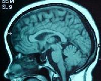
empty sella-MRI.sag
|
|
Sometimes
bilateral and symmetrical lateral shift of the carotid artery can be
seen, or descent of the initial portion of the anterior cerebral artery
into the empty sella turcica occurs occasionally.
Treatment:
Primary
as well as secondary empty sella syndrome is usually benign conditions
not requiring any treatment.
However,
in some cases the clinical and endocrine status necessitates hormonal
replacement therapy. Evidence of pituitary hypersecretion indicates
the presence of a hypersecreting adenoma or remnant or recurrent tumor
after surgical resection or radiotherapy that should be treated
accordingly.
Visual
field disturbances usually are mild and should therefore only be followed
carefully. Surgery should be reserved for cases with progressive
deterioration and radiological evidence of traction or angulation of the
optic nerves and chiasm.
The
surgical process of propping up the optic chiasm was called chiasmopexy.
Several methods are described with good results:
a)
Inserting muscle, carilage, or silicone sponge under the optic chiasm,
b)
Packing the sella with fat, muscle, or cartilage to elevate the pituitary
gland, pituitary stalk and chiasm via the transsphenoidal route,
c)
Packing the sella by transsphenoidal placement of a detachable balloon
(Cybulsky 1989),
d)
Mortata (1970) demonstrated improvement of visual field defects in a
patient with empty enlarged sella and descent of
CSF
rhinorrhoea may require prompt surgical intervention since spontaneous
obliteration is unlikely. After appropriate preoperative
localization of the fistulous tract (fluorescein) endoscopic or
transsphenoidal closure is the treatment of choice.
|
