|
Spinal disorders
and the pain associated with them account for a significant portion of
disability at work, with most complaints occurring in the lumbar region.
Men are more frequently affected. It is essentially a disease of the
middle aged, unless precipitated by trauma. L4/5 and L5/S1 disc
account for 90% of the cases, with each level affected about equally.
L3/4 disc accounts for the majority of the remaining herniations.
Symptoms:
In the acute presentation, symptoms often follow
trauma or an injury to the disc produced by a sudden spinal strain, such
as lifting heavy weights. There is acute low back pain, and, in the event
of nerve root compression, radiating pain, paresthesias, and motor
weakness. Severe bilateral root dysfunction may produce bowel and bladder
incontinence and sexual dysfunction. If the leg pain is not immediately
experienced, it usually appears over ensuing hours with associated
paresthesias.
The leg pain is usually worse and in dermatomal fashion. The
back pain is thought to be secondary to activation of the sinu-vertebral
nerves to the annulus, which share a central pathway with the nerve
roots.
Chronic form is characterized by chronic
intermittent exacerbations of back ache, usually without leg pain
initially. The backache usually subsides with rest and conservative
measures, only to reoccur. The leg pain may be as well localized as in
the acute form.
The pain , unlike the pain due to tumors, is usually
subsides to some extent and with recumbency; the pain is aggravated by
increased activities, bending forwards, coughing, sneezing, and straining
at stools. Prolonged sitting increases the intradiscal pressure and the
pain.
Signs:
On examination, the patient presents with findings of axial
pain and radiculopathy.
a) The paraspinal muscles are often in spasm, particularly
on the side opposite the leg pain, and are tender to palpation.
The patient leans away from the side of leg pain with the hip and knee
flexed in an effort to reduce the leg pain.
Spinal movements are restricted due to pain.
Lateral bending towards the side of leg pain closes the
intervertebral foramen, compressing the nerve root, and worsens the pain.
The straight leg raising test and the crossed straight leg (
contra lateral leg) raising stretches the sciatic nerve and L5 and S1
root pain get worse.
The reverse straight leg (femoral stretch test) raising
stretches the femoral nerve and reproduce the leg pain in the
distribution of the affected nerve root contributing to the femoral
nerve(L2,3 or 4).
b) Neurological examination may detect the motor, sensory,
and reflex impairment (LMN type).
In the lumbar region, the nerve roots exit through the
intervertebral foramen caudal to their corresponding vertebral pedicle
(eg: the L5 root exits at L5/S1 intervertebral foramen); the majority of
disc herniations, being posterolateral, compress the root that exits at
the intervertebral foramen below the level of the involved disc (L5 root in
L4/5/ posterolateral disc herniation). In the less commoner lateral
herniation, the root, that exits the foramen at the level of the involved
disc, is affected (L5 root in L5/S1 lateral disc herniation).
L5 radiculopathy may present with pain, and paresthesia/numbness
along the posterolateral aspect of the leg down to the great toe;
weakness of extensor hallusis longus and dorsiflexion may be noted.
S1 radiculopathy may present with pain, and
paresthesia/numbness along the posterior aspect of the leg down to the
lateral aspect of the heel and foot; weakness of ankle plantar flexion
may be detected. Ankle jerk may be absent.
L4 radiculopathy (as in the posterolateral disc herniation
at L3/4) may present with pain and paresthesia/numbness along the
anterolateral thigh and below the knee to the medial aspect the leg and
foot; weakness of quadriceps and knee extension may be noted.
L3 radiculopathy the pain and paresthesia may be localized
over the anteromedial thigh and in L2 the distribution is over the groin.
Both L2 and L3 radiculopathies may cause quadriceps weakness.
Large central discs may cause bilateral symptoms or cauda
equina syndrome. characterized by asymmetric pain and paresthesias in the
perineum and down the back or front of the thighs and legs. Sensory
disturbances may be limited to the backs of the thighs, buttocks, anus,
and perineum, so called saddle anesthesia. paralysis of the bladder and
rectum with associated incontinence may occur if the compression is low
and affects the sacral roots bilaterally.
|
Diagnosis:
MRI is the test of choice for evaluation of disc disease.
its multiplanar capabilities make it suitable for visualizing far
lateral disc herniations as well as the paravertebral structures.
CT myelography still is useful in patients with equivocal
MRI studies and in those who are unable to undergo MRI scanning.
Management:
Medical:
|
|
|
|
The initial
management is nonoperative, unless the patient presents with
significant neurological deficit.
|
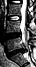
L5/S1 disc prolapse-
MRI
|
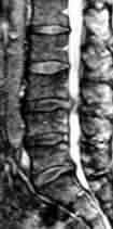
L3/4 disc prolapse-
MRI
|
The medical management traditionally involves bed rest and
analgesics and anti-inflammatory drugs. Muscle relaxants help in some.
TENS helps in about 20% of patients.
Surgical:
Indications for surgery include failure of acceptable pain
control by nonoperative measures, progressive neurological deficit, and
cauda equina syndrome.
The traditional approach to lumbar discectomy is through
posterior hemilaminotomy and foraminotomy, either in prone position or
'knee-elbow' position, usually under general anesthesia. Use of a microscope
helps and has become a routine these days.
Lately, fenestration (microlumbar discectomy
- wherein no bone is removed and the disc is approached by excising the
ligamentum flavum at the required level) is increasingly employed with
good results. The advantages in addition to minimal disturbance to
spines, are less post operative discomfort and less stay in the hospital.
Whichever approach is used, at least 10gms of disc material
has to be removed for adequate pain relief.
The 'so-called' minimally invasive procedures, such as
percutaneous chymopapain injection, automated percutaneous discectomy,
percutaneous laser discectomy, and endoscopic discectomy are being
advocated by some with varying results. Among these, endoscopic
discectomy has the potential to become the choice of treatment in future.
Complications of surgery:
Bacterial discitis is a most often considered
complication, especially when there is recurrence of pain and tender
spines. It may warrant a corpectomy and fusion.
Dural injury must be avoided; valsalva manoeuvre and primary
closure of the dural rent at the end of discectomy is preferable. Failure
to close the rent will produce pseudomeningocele and recurrence of
symptoms. CSF fistula may result and needs to be repaired.
Extensive epidural scarring may present with recurrence and
'failed-back' syndrome. Treatment is medical.
Life threatening injury to the major vessels, such as aorta,
vena cava, and iliac vessels have been reported. Bowel perforation may
also occur.
Incidence of recurrent disc herniation is thought to be
about 10% and about 1/3rd of them in the first year. They are treated the
same way as at the original episode. Fusion may be required in some
patients, and recommended as a routine by some.
Outcome:
Approximately, 2/3 of the patients with acute sciatica
recover within 4 weeks; about 1/3 of them report with recurrence.
The main advantage of surgery is to accelerate the time to
recovery. In most studies, there is a definite advantage to surgery in
the first postoperative year. In general, patient selection is important.
A motivated patient, with history, physical findings, and diagnostic
studies confirming nerve root compression, can be expected to do well.
90% of the operated patients do well.
Patients with chronic pain, and on prolonged use of narcotic
analgesics, and patients with psychological dysfunction orpending
litigation for compensation should alert the surgeon of possible poor
outcome.
|
