|
With an increased knowledge of the surgical anatomy and
multidisciplinary approach, most of the cranial basal lesions can be
dealt with safely. Newer techniques in reconstruction of blood
vessels and nerves, sophisticated neuro anesthesia and
intensive post operative nursing are of great help. It is possible to
keep the brain retraction to the minimum and damage to the blood
vessels and nerves are avoided with newer skull base techniques.
The aim is to get the extra room under the brain.
SURGICAL ANATOMY:
A) Osteology:
1) The body of the sphenoid occupies the central
portion. The tuberculum sellae is a transverse ridge , that separates
the chiasmal sulcus anteriorly from the sella turcica posteriorly.
The sella is a rounded hollow that cradles the pituitary gland.
2) The sides of the body slop down and
laterally, grooved by the sigmoid curve of the ICA, to the floor of
the middle fossa.The anterior and posterior clinoid processes are
important landmarks and areas of dural attachment .Occasionally there
is a middle clinoid process, that may be bridged to the anterior
clinoid, so forming a caroticoclinoid foramen through which passes
the ICA.
3) The lateral recesses are the middle fossae
proper and triangle shaped, limited anteriorly by the spenoid
ridge and posteriorly by the petrous ridge.
-The anterior
wall is formed by the greater wing of the sphenoid.
-The floor is by
the greater wing anteriorly, and the petrous ridge posteriorly.
Laterally, between the two is the squamous temporal
bone.
-Thelateral wall
is made up of the greater wing of the spenoid anteriorly and the
squamous temporal bone posteriorly.
-The posterior wall
is by the petrous ridge.
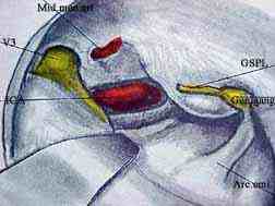
The floor and lateral walls are grooved by the
middle meningeal artery. The superior surface of the petrous ridge
has several important markings.
-Medially, near the apex
is an impression for the trigeminal ganglion as it lies in the
Meckel's cave. The ICA runs directly under this and the bony canal
may be dehiscent.
-Laterally is the thin
tegmen tympani, roofing the middle ear and mastoid.
- Anteromedially, lies
the arcuate eminence overlying the superior semicircular canal.
Further antero-medially, lie the canals for the greater and lesser
superficial petrosal nerves. GSPN may be traced to the
geniculate ganglion and facial nerve in the IAC.The bone may be
dehiscent over the geniculate ganglion.
-The petrous ridge is
longitudinally grooved by the superior petrosal sinus where the
tentorium cerebelli attaches.
4) The foraminae:
-Anteriorly lies the
superior orbital fissure, which leads to the orbital apex.
-Foramen rotundum lies
behind and inferior to superior orbital fissure and transmits the
maxillary division of the trigeminal nerve.
-Foramen ovale lies
posterolateral to the foramen rotundum and transmits the mandibular
division of the trigeminal nerve, the accessory meningeal artery, the
lesser superficial petrosal nerve and emissary veins to the pterygoid
plexus.
-The foramen spinosum
lies posterolateral to the foramen ovale and transmits the middle
meningeal artery.
-The petrous apex
articulate with the sphenoid and occipital bone medially and so forms
a rounded opening to the carotid canal (cranial counterpart of the
foramen lacerum) on the under surface of the skull base.
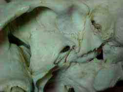
|
|
1-optic canal
2-superior orbital fissure
3-For.rotundam
4-Venous For.
5-For. ovale
6-For.spinosum
7-For. lacerum
8-Groove for GSPN
9-Groove for mid.men.art
|
|
|
5) The temporal bone itself contains several
important structures.
-The sigmoid sinus ends
in the jugular bulb.
-The 7th and 8th nerves
enter the porus-acusticus and IAC. The 7th nerve traverses the
middle ear and mastoid. The 8th nerve ends at the inner ear.
-The eustachian tube
arises at the protympanum and runs anteromedially and inferiorly
.The tube is one third bony and two thirds
cartilaginous.
-Directly medial to the
origin of the bony eustachian tube lies the ICA.
|
|
|
B) Intracranial contents:
1 ) The dural arrangement is complex and
densely adherent in the regions of clinoid processes, petrous
and sphenoid ridges and around the basal foraminae. In the midline it
forms a transverse dural plate, the diapraghma selle, roofing the
pituitary fossa. Laterally the dural plate forms the roof of a basin
beside the body of the sphenoid,the cavernous sinus.
2) The cavernous sinus is a plexus of veins
that lies within the layers of the dura beside the sphenoid sinus.
The lateral border of the roof is the anterior petroclinoid fold and
the posterior border is the posterior petroclinoid fold.
The ICA is the main structure within the sinus. The 6th
nerve is the only nerve within and lies in close opposition to the
lateral wall of the ICA. The cranial nerves the 3rd, 4th, and 5th are
variably related to each other in the lateral wall.
Parkinson has outlined triangles between these nerves
that can be used to gain access safely to the cavernous sinus.
Anterior venous connections are the superior ophthalmic
vein and spheno-parietal sinus. Superiorly, the cavernous sinus
drains the superficial middle cerebral and inferior cerebral veins.
Medially is the intercavernous plexus to form the circular sinus.
Inferiorly, the emissary veins pass to the pterygoid plexus.
Posteriorly it drains into the superior and inferior petrosal sinuses
and into the basilar plexus between the dural layers over the clivus.
3) The motor and sensory roots of the 5th nerve
pass underneath the free edge of the tentorium cerebelli and into the
Meckel's cave, which contains the motor root and trigeminal
(Gasserian) ganglion, which overlies the petrous apex and ICA.
The ganglion is variably enclosed by the subarchnoid space and
CSF. The cranial nerves V1, V2 and V3 pass from the ganglion into the
lateral wall of the cavernous sinuses. The motor root passes
with V3 through the foramen ovale.
4) The temporal lobe fills most of the rest
of the fossa.
5) The inferior anastomatic vein (of LABBE)
connects the superficial middle cerebral vein to the transverse sinus
just before it becomes the sigmoid sinus. Injury to this vein may
result in infarction of the motor cortex. The superior anastomatic
vein (of TROLARD) connects the middle cerebral
vein to the superior cerebral veins.
6) The greater petrosal nerve (GSPN) and
the lesser petrosal nerve (LSPN) run parallely beneath the
dura along the anterior edge of the petrous bone as it runs to the
foramen lacerum .It is also a landmark for the ICA which lies just
deep and parallel to it.
7) The internal carotid artery is the most
important structure at risk during surgery.
It is divided into four parts:
-The cervical
portion arises at the 3rd and 4th cervical vertebrae, runs superiorly
to the external carotid artery and deep to the digastric muscle
and styloid apparatus. The glenoid fossa is a bony landmark for the
higher parts of the ICA at the eustachian tube level.
This portion
has no branches.
-The intra temporal
ICA has a vertical and a horizontal segment: -The vertical segment
(C1) begins at the canal where it is anchored very
firmly by a fibrinous ring. It ascends for 5mm, turns anteromedially
into the horizontal segment(C2) which runs forward in
the petrous bone directly related antero-laterally to the eustachian
tube in this portion.
-The cavernous portion of
the ICA ( C3 ) is very thin walled.
-The supracavernous ( C4 )
portion begins as the artery pierces the dura in the roof of the
cavernous sinus medial to the anterior clinoid process, passes
backward below the optic nerve to the anterior perforated substance
where it in the circle of Willis.
|
C) The Infra-temporal fossa:
It is the undersurface of the middle cranial
fossa.
The bulk of it, is occupied by the
lateral and medial pterygoid muscles. Intimately related, are
the branches of V3, the pterygoid plexus of veins and branches of
the maxillary arteries. Deeper, arising from the skull base and
cartilaginous eustachian tube, are the tensor and levator veli
palatini muscles. At the deepest, most anterior part of the
infratemporal fossa, lies the pterygoid process and more anteriorly
,the pterygomaxillary fissure ,leading into the pterygomaxillary
fossa.
Medially, the sphenoid sinus
lies anteriorly and the nasopharynx posteriorly. More
posteriorly, is the clivus. Directly above the nasopharynx is the
foramen lacerum, plugged by fibrous tissue and cartilage, and
directly above this, lies the carotid in its canal just before it
enters into cavernous sinus. The gap between the superior
constrictor of the nasopharynx and skull is the foramen of Morgagni
which is largely filled by the eustachian tube and palati muscle.
It is a potential route for tumor spread.
Laterally lies the parotid
gland and facial nerve, then the zygomatic arch and mandibular
condyle. The temporalis muscle inserts onto the coronoid process of
the mandible with temporal arteries on its undersurface, which
needs to be preserved so that the muscle can be used in
reconstruction.
Immediately posterior to the styloid process, lies the
stylomastoid foramen, where the facial nerve exits. Directly
poterolateral lies the jugular foramen, where the 9th, 10th and
11th cranial nerves become intimately related to the great
vessels.Posteromedial to the carotid canal, lies the occipital
condyle and under its tip, the hypoglossal canal where the 12th cranial
nerve exits.
|
|
Drilling medially through the glenoid fossa leads
straight into the bony eustachian tube and superiorly, into the
middle cranial fossa. Anteromedially is the foramen spinosum , and
then the foramen ovale.
Further medially, is the eustachian tube (cartilaginous)
and more medially, the carotid artery.
SURGERY:
Preoperative work-up:
1) MRI with and without gadolinium is valuable
.Fine sections reveals encasement of the blood and suggest the nature
of the lesion.
2) CT with bone windows shows the bony changes.
3) Cerebral angiography reveals the
vasculature of the lesion and cross circulation. Pre operative
embolization may be required in selected cases.
4) Discussion with the ENT surgeon, the
Anesthetist and scrub nurse regarding the objective of the procedure,
whether radical excision or otherwise, and positioning, CSF
drainage during the procedure etc, is a must.
5) Neurophysiological monitoring, if available,
may be useful.
Approaches:
The sphenoid-wing meningiomas, sphenocavernous and
cavernous lesions, tumors of and around The petrous bone and some
complex and giant aneurysms are better dealt with skull base
approach. Pituitary adenomas and Craniopharyngiomas with para-sellar
extension may require this route for radical excision. Tumors from
the infratemporal fossa may also extend into the intracranial cavity.
With added modifications, the middle fossa approaches may be employed
for lesions around the lower clivus as well.
The extent of the lesion, objective of the surgery,
availability of the facilities and experience of the surgeon in
skull-base surgery decides the approach, as in any surgery of any
kind.
1)Anterolateral approach:
This approach is recommended for lesions around
and above the level of the upper clivus, above the level of the
crossing of the 5th and 6th nerves from posterior to middle fossa.
--Under general anesthesia, the patient is
positioned with the head turned to the opposite side.
--The common and internal carotids are exposed at
the neck for future temporary occlusion.
--Through a bicoronal (if a frontobasal approach
is also planned) or a frontotemporal scalp incision, a frontotemporal
craniotomy is made .I prefer a free bone flap.
|
--The next step is the orbitozygomatic
osteotomy.
-A cut is made in the
sagittal plane at the medial aspect of the orbit across the
superior rim and wall of the orbit at or near the supraorbital
notch and extending about 2.5cm posteriorly.
-A second cut is made
in the coronal plane across the orbital roof and then across
the lateral wall of the orbit to the inferior orbital fissure.
|
|
|
The
area of Orbitozygomatic osteotomy
|
-The anterior zygomatic osteotomy is made at or lateral
to the zygomaticomaxillary suture.
-The posterior zygomatic
osteotomy is then made through the condylar fossa or just anterior.
-The entire orbital rim,
the zygomatic arch and condylar fossa may then be removed as a single
piece.
--Next, the drilling of the sphenoid wing is done
untill the base of the clinoid.Ideally the ant.clinoid is removed
extradurally, some prefer to remove the anterior clinoid
intradurally to prevent injury to the surrounding structures.
--CSF drainage at this stage helps.
--V2 and V3 branches are exposed extradurally in
the subtemporal area, and the superior orbital fissure is
decompressed.
--The dura-pericranial hitch stiches are applied.
The dura is opened and turned anteroinferiorly as a flap.
--Another slit in the dura along the sylvian
fissure provides a protective cover to the frontal and
temporal cortex
--The sylvian fissure is opened laterally and the
branches of the middle cerebral artery are followed proximally
--The optic canal is decompressed. The optic nerve
sheath is opened to mobilize the nerve. The dural rings around the
ICA are opened.
--The tumor is removed in piecemeal using
microsurgical techniques. It is prudent to leave behind the
tumor bits adherent to vital structures.
--If necessary, the cavernous sinus is opened at a
point where the lesion presents as a bulge or through one of the
parkinson's triangle and the dural layer is peeled away. The 3rd,
4th and 5th nerves are at risk and must be protected at this stage.
In the presence of a lesion, the venous plexus is collapsed, and
bleeding is not a problem. The dissection of the 6th nerve can be difficult
and must be done carefully. Some bits of the lesion may have to be
left attached to the ICA. Small tears in the artery may need to
be sutured after temporary occlusion of the carotid at the
neck. When the artery is completely encased, excision of the
involved, the ICA may be contemplated with a vein graft bypass. Some
prefer ICA or ECA to MCA bypass. Many leave the adherent tumor
behind.
When direct surgery is planned, intracavernous giant
aneurysms are dealt with, after temporary occlusion of the carotid at
the neck.
Induced hypertension, mild hypothermia and
barbiturate coma are used during vascular occlusion.
--The clival and sphenoidal bone may need to be
drilled on occasions for complete tumour removal.
--The tentorium overlying the Meckel's cave may be
opened, exposing the prepontine and interpeduncular area, to access
into the posterior fossa, if required.
--Following excision, the cavernous sinus is
repaired with fascia lata. If the sphenoid sinus is entered, it
is packed with fat and the dura is closed watertight.
--The orbito-zygomatic arch and then the bone flap
are replaced, followed by the scalp closure.
|
2) Subtemporal-infratemporal approach:
This approach is recommended for the tumors involving the
petrous and sphenoid bone and gives access to entire mid clivus,
down to the level of the 11th nerve.
It is also useful for tumors involving the
infratemporal fossa and ptrygopalatine fossa.
--Under general anesthesia, the patient is
placed in the lateral position.
--The upper cervical carotid may be exposed and
kept secured for future temporary occlusion.
--A bicoronal incision with preauricular
extension is made. Below the zygomatic arch the dissection
are kept close to the ear, preserving the superficial temporal
artery, keeping the dissection plane just superficial to the
massetric fascia to avoid injury to the facial nerve. The massetric
fascia and muscle are detached from the zygomatic arch.
--Depending on the extent of the tumor, a
temporal craniectomy or a frontotemporal craniectomy is
performed, extending to just above the mastoid process posteriorly.
--Next, a orbitozygomatic osteotomy or
zygomatic osteotomy including condylar fossa is performed. If more
posterior room is needed, the condyle and condylar fossa are
included. The temporomandibular joint capsule is opened, the
meniscus is dissected and depressed. The attachment of pterygoid
muscles must be divided. The styloid process is a landmark. The
dissection should not go deeper at this point.
--V2 and V3 branches are exposed extradurally and the
superior orbital fissure is decompressed.
--Next is the mobilization of petrous ICA.
|
|
|
|
Extradural dissection -1
|
|
|
|
|
Extradural dissection -2
|
|
|
|
Extradural dissection -3
|
|
The petrous ICA is often partially exposed without any
bony covering just posterior and medial to V3 and middle meningeal artery
and inferior to the GSPN.
The bone between the middle cranial fossa and
mandibular fossa may be removed to expose the genu of ICA.
Care must be taken not to injure the cochlea or the
geniculate ganglion and the facial nerve which lie immediately posterior
and superior to the genu.
The ICA is identified in one area, the entire ICA
is progressively exposed and unroofed.
The bone medial to V3 and lateral to ICA may have to be
drilled just medial to V3.The lumen of the eustachian tube
is cauterized and packed with muscle and fat and closed.
The jugular bulb and cranial nerves 9,10 and 11 lie
immediately posterior to the vertical segment of the petrous ICA.
The petrous apex medial to ICA can be progressively
removed and the midclival and petrous apex dura can be exposed.
Medial to the vertical ICA, progressive removal of the
bone will allow unroofing of the 12th nerve.
Now, the entire petrous and upper cervical ICA is
exposed and mobilized.
--The sphenoid sinus is approached anteriorly
between V2 and V3.
--The tumor is removed in piecemeal using
microsurgical techniques.
--The cavernous sinus may be entered extradurally
or intradurally to complete the tumor removal with an
appropriate dural incision.
--The V3 may be divided to access the lower
clivus, sphenoid and opposite petrous apex.
--The defects are closed with an autologous fascia
lata graft.
The dead space is filled by a vascularized
temporalis muscle flap or a distant microvascular free
flap.
--A post-operative CSF drainage is often employed
to prevent a CSF leak.
Many patients require some type of
rehabilitation for ocular, facial, swallowing and speech
disorders postoperatively.
|
