|
Spinal metastases
are almost always diagnosed after the diagnosis of the primary cancer.
25% of spinal tumors in children are metastatic deposits. 80% of those
with bone metastases will have a spinal involvement. They are more
frequent in the elderly (6th and 7th decade). There is a slight male
preponderance.
Pathophysiology:
Primary sources for spinal metastatic disease include the
following: Lung (31%), Breast (24%), GI (9%), Prostate
(8%), Lymphoma (6%), Melanoma (4%), Unknown (2%),
Kidney (1%), Others including multiple myeloma (13%). Time relation
between primary and spinal metastases vary according to the site and
nature of the primary.
Spread from primary tumors is mainly by the arterial route
via nutrient artery. Retrograde spread through the Batson plexus during
Valsalva maneuver has been postulated. Direct invasion through the
intervertebral foramina also can occur.
About 70% of symptomatic lesions are found in the thoracic
spinal region, 20% in the lumbar region, and 10% in the cervical spine.
Over 50% of patients with spinal metastasis have multiple level
involvement. About 10-38% of patients have multiple noncontiguous segment
involvement. Most of the lesions are localized at the anterior portion of
the vertebral body (60%). In 30% of cases the lesion infiltrates the
pedicle or lamina. A small percentage of patients have disease in both
posterior and anterior parts of the spine.
Intramural and intramedullary metastases are not as common as
those of the vertebral body and the epidural space. Isolated epidural
involvement accounts for less than 10% of cases; it is particularly
common in lymphoma and renal cell carcinoma. Epidural
metastasis is the most ominous complication of bone metastasis to the
vertebral spine and is a medical emergency. The tumor enters the-epidural
space by contiguous spread from adjacent vertebral metastasis in the vast
majority of cases. The remaining cases arise from the direct invasion of
retroperitoneal tumor or tumor located in the posterior thorax through
adjacent intervertebral foramina or, rarely, from bloodborne seeding of
the epidural space.
Besides mass effect, an epidural mass can cause cord
distortion, resulting in demyelination or axonal destruction. Vascular
compromise produces venous congestion and vasogenic edema of the spinal
cord, resulting in venous infarction and hemorrhage. The relative
importance of vascular factors as opposed to purely mechanical ones has
been a subject of controversy for many years. The tempo of development of
spinal compression is, perhaps, impossible to generalize. Once
neurological symptoms become manifest, the condition is a neurological
emergency.
Clinical
presentation:
Bone pain at night in a patient with cancer is always an
ominous symptom. The majority of the patients present with radicular
pain. The
pain is usually midline, but patients whose tumor involves nerve roots
have sharp or shooting pain in a radicular distribution. Untreated, the
pain slowly intensifies with a mean duration of 7 weeks from the onset of
pain to the onset of neurological deficits due to spinal cord
compression. Half of these patients have
sensory and motor dysfunction and over 50% have bowel and bladder dysfunction.
About 5-10% of patients with cancer present with cord
compression as their initial symptom. Among those who present with cord
compression, 50% are nonambulatory at diagnosis and 15% are paraplegic.
Diagnosis:
|
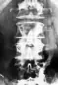
Plain x-ray is used to show erosion of the pedicles
or the vertebral body. More
than 70 percent of patients with spinal cord compression have an
abnormal plain radiograph in the region of pain (compression fracture,
plastic, or lyric metastasis). Owl eye
erosion of the pedicles in the anteroposterior (AP) view of lumbar
spine is characteristic of metastatic disease and is observed in 90% of
symptomatic patients. Osteoblastic or osteosclerotic changes are common
in prostate cancer and Hodgkin disease; occasionally, they also are
seen in breast cancer and lymphoma.
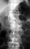
CT scan is useful in determining
the integrity of the vertebral column, especially when surgery is
anticipated. CT myelogram is
|
|
|
used if
MRI is not available.
|
|
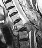
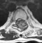
MRI scan is the choice of imaging. MRI sagittal
scout film is used for rapid screening of the surrounding soft tissues. Patients with
persistent back pain in the region of abnormality on plain spine
radiograph, with or without neurological deficits, should undergo
evaluation with MRI. Patients with progressive back or neck pain whose
plain radiograph is normal should also undergo an imaging study of the
epidural space, even if their neurological examination is normal.
|
|
|
|
|
|
|
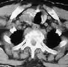
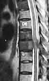
D2 met-MRI-sag
|
D2 met-MRI axial
|
D2 -met -CT
|
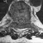
D8 hyperostotic met-MRI sag
|
D8 hyperostotic met-MRI
axial
|
Emergency myelogram still is used in situations where
MRI is not available. Myelograms, allow for cerebrospinal fluid (CSF)
sampling. CSF sampling should be deferred if evidence of near/complete
spinal block is noted. The risk of having neurological deterioration
after myelogram is about 14%. Neurological deterioration is less likely
with C1-2 puncture.
Bone scan findings are positive in
60% of cases.
Needle
or open biopsy will establish the diagnosis.
A thorough metastatic workup is paramount in patients
with spinal metastasis. This helps to delineate the nature and the
extensiveness of the systemic disease; however, the appropriateness of
diagnostic tests depends on the amount of time available. In patients
with rapidly progressing symptoms, chest x-ray and physical examination
is all that is permitted. The patient should then have a plain x-ray of
the entire spine, followed by MRI with and without contrast.
Management:
Medical therapy: Immediate treatment is high-dose
dexamethasone and analgesics. The optimal dexamethasone dose has
not been established, but in practice the usual dose is 4 mg hourly after
a loading dose of 24mg. Of all the corticosteroids, dexamethasone has the
least mineralocorticoid effect and is least likely to be associated with
infection or cognitive dysfunction, although it does increase the risk of
myopathy. The frequency of complications from steroid therapy is
dependent on the duration of the treatment and is associated with
hypoalbuminemia. Treatment lasting more than 3 weeks is more likely to be
associated with complications. Hypoalbuminemia appears to increase the
risks of adverse effects associated with steroid treatment.
About 70-80% of patients experience improvement of symptoms
within 48 hours of treatment. Approximately 64% of patients report
alleviation of pain within 24-48 hours of starting steroid therapy and
57% show improvement in their motor function. In most cases steroid use
needs to be continued until the completion of radiotherapy.
Radiotherapy: Radiotherapy remains the mainstay
of treatment for spinal metastatic disease. Most of the lymphoreticular
tumors and prostate carcinoma are radiosensitive; lung and breast are
less sensitive. Tumors of the gastrointestinal system and kidney are
resistant to radiotherapy, as are melanomas.
Nevertheless, radiotherapy has been offered to the latter
group of patients and has demonstrated some response. The radiation port
normally includes 2 vertebral bodies above and below the diseased
segment. 1000-1200 rads a week in divided doses for 3-4 weeks, for an
average total of 3200 rads is the usual dose. About 80% of patients with
pretreatment pain have symptomatic relief; 48% of patients with motor or
sphincter dysfunction respond to treatment.
Surgical therapy: Most often surgery
is indicated only as a stabilization procedure or for tissue diagnosis.
It is employed in patients who have disease progression despite
radiotherapy and in those with known radiotherapy-resistant tumors.
The
patient should be suitable for adjuvant therapy.
The
results of surgery are superior to those of irradiation alone. Relief of
pain and restoration of neurological deficit are respectively 82% and 70%
after the surgery, and 56% and 55% after the irradiation. The duration of
survival following surgery tended to be related to primary tumor. Minimal
survival is achieved in lung carcinoma, and maximum survival is achieved
with prostate carcinoma. Although the surgery of bone metastasis does not
necessarily affect the life expectancies of the patients, adequate
surgery is often able to provide a patient with several years of
pain-free, mobile and useful life.
Neurological
deficit and back pain are caused not only by cord or root compression,
but also by skeletal instability. Therefore, the surgical results after
the decompression and stabilization were superior to those after the
decompression alone when they were evaluated on the basis of pain relief
and restoration of neurological deficit.
|
The selection of a
surgical approach is influenced by the primary site and extent of
osseous and neural involvement and the patient's preoperative
neurological and clinical status.
The
current trend is to decompress anteriorly (vertebrectomy, corpectomy)
for anteriorly placed lesions. It is always combined with
reconstruction and and stabilization with instrumentation.
Laminectomy is indicated less commonly than these other
procedures, because most of the lesions are anteriorly based, and
posterior decompression may further destabilize the spine.
In
a potentially curable lesion, a combined anterior and posterior
decompression may be indicated.
|
|

|
|
|
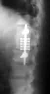
Titanium cage & compression screws- AP
|
Titanium cage & compression screws- Lat
|
|
|
|
|
Stabilization with instrumentation
|
|
Surgical decompression and stabilization together with
radiotherapy is promising.
This offers stabilization of the diseased bone and allows
ambulation together with pain relief. Patients with nonambulatory status
at diagnosis do poorly, as do patients with more than one vertebra
involved.
In
selected cases, chemotherapy helps as an adjuvant to surgery and
radiation. Hormone therapy, as in secondaries due to prostatic
carcinomas, give good palliation.
Conclusion:
The outcome of metastatic disease to the spine and associated
structures is uniformly bleak.
The median survival duration for patients with spinal
metastatic disease is 10 months.
The morbidity of spinal metastatic disease is of
significance, especially in patients with paralysis and/or bowel and
bladder involvement. The latter compromises the quality of life of
patients with cancer and puts an additional burden on the caregiver.
The ultimate goals are to maintain the independence and
dignity of the patient and to optimize his or her comfort level.
No treatment has been proven to increase the life expectancy
of patients with spinal metastasis. The goals of therapy are pain control
and functional preservation. The most important prognostic indicator for
spinal metastases is the initial functional score. The ability to
ambulate at the time of presentation is a favorable prognostic sign. Loss
of sphincter control is a poor prognostic feature and is mostly
irreversible.
Radiation therapy is more effective in achieving pain
control (67%) than surgery (36%). Notably, surgery alone is the least
effective way to treat spinal metastases. About 20-26% of patients who
undergo surgery experience further deterioration in terms of either
mobility or sphincter control while only 17% in the radiation therapy
group experience further deterioration.
Surgical intervention with extensive reconstruction should
be employed only after thorough evaluation of the extent of the systemic
disease and with a clear understanding of the realistic expectation of
the patients and their caretakers.
|