|
Spinal
tuberculosis is common in the developing countries and also seen
sporadically in well-developed countries. Lately the incidence is on the
increase, world over, with the emergence of AIDS. About 60 % of cases are
below the age of 20 years in developing countries. In developed countries
the older people are more commonly affected.
About 20% of the
patients have multiple lesions.
Most are caused by the human strain. The bovine type is
probably responsible for less than 5 %, especially in Europe. Isolated
cases due to atypical mycobacteria are also seen.
|
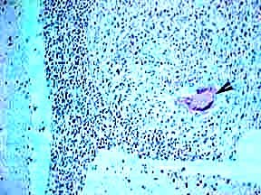
Pathology:
Microscopically, there is central coagulative necrosis
surrounded by epitheloid cells, Langhans giant cells(as shown by the arrow
in the picture) and an admixture of lymphocytes and plasma cells. There
may be satellite lesions and perivascular infiltrations
Tuberculosis may involve the vertebra, epidural space,
dura, arachnoids, or spinal cord.
A) Vertebral involvement:
|
|
It is the commonest. It is also the commonest form of
skeletal tuberculosis with an incidence of up to 50% of all skeletal
tuberculosis. In general it is a disease of the young adult in the
developing countries. In developed countries it affects more commonly,
the elderly. Due to the emergence of HIV infection the incidence of all
forms of tuberculosis is further aggravated all over the world. Both
sexes are equally effected.
The spinal disease is always secondary to a primary lesion
and occurs due to hematogenous spread. The primary focus may be active or
quiescent and may be in the lungs, mediastinal lymph nodes, kidneys and
other viscera. On an average, an involvement of 3 - 4 vertebrae at the
time of presentation has been reported. As elsewhere, the spinal
tuberculosis is a granulomatous disease. Marked exudative reaction is a
common feature of spinal tuberculosis. A cold abscess mostly comprised of
serum , leucocytes, caseous material, bone debris and bacilli, penetrates
the ligaments and migrates along the facial planes often presenting far
from the site of infection.
Clinically there are four types :
1. Para discal lesion begins in the metaphysis, erodes the
cartilage and destroys the disc, resulting in narrowing of the disc
space.
2. Central type begins in the midsection of the body which
gets softened and yields under gravity and muscle action, leading to
compression, collapse and bony deformation.
3. Anterior lesions lead to cortical bone destruction
beneath the anterior longitudinal ligament. Spread of the infection is in
the subperiosteal and sub ligamentous planes resulting in the loss of
periosteal blood supply to the body with resultant collapse. Other
factors such as periarteritis and endarteritis contribute to the
collapses.
4. In appendicle type, the infection settles in the
pedicles, the laminae, the articular processes or the spinous processes
and causes initial ballooning of the structure followed by destruction.
Tuberculous spondylitis commonly occurs in the thoracic,
followed by lumbar and cervical spines which more often occurs in the
pediatrics group.
Clinical features:
1. Back pain is a predominate (70%) feature with stiff spine
and Para vertebral muscle spasm. A soft tissue swelling or mass is often
obvious. There is 20% incidence of cold abscess and about 90% incidence
of angulations of the spine in the form of kyphosis or gibbus.
2. Systemic symptoms may or may not be there.
3. The most serious is the neurological involvement with
overall incidence of about 30% and the deficit depends on the site, the
direction of spread and pathological changes produced. The risk is
highest in the region of cervico-thoracic region.
The cord may be involved in any phase, the active phase
within the first 2 years or in later years after the disease has become
quiescent. The cause in most cases is compression, which may be an
abscess, granulation tissue, sequestrated bone and disc or pathological
subluxation in active disease.
In healed diseases the deficit may be due to transverse
ridge of bone anterior to the cord, due to angulations of the spine or
healing, stretching or attrition of the cord due to spinal deformity and
or fibrosis of the dura.
In a given case more than one factor may contribute to the
pathogenesis. Non compressive causes such as endarteritis, periarteritis
or thrombosis of the arterial supply of the cord.
As mentioned earlier, cervical spine involvement is rare
(1%) more often seen in children. It is characterized by a more diffuse
involvement of the lower cervical spines the formation of
retropharyhngeal abscesses, often causing respiratory distress. The adult
form is usually confined to a single body and more commonly results in
kyphosis and cord compression.
TB of CV may cause atlanto axial subluxation, upward
translocation of the dens, cervico medullary compression of tuberculous
abscess or direct invasion by the disease. The disease infiltrates the
ligaments which give way. Incidence of associated lesions vary between 40
- 50%.Simultaneous involvement of other bones has been reported to be
between 12-15%.
Diagnosis:
Suspicion is the first step in diagnosis. No diagnostic
procedure either singly or in combination will provide an unequivocal
diagnosis.
The erythrocyte sedimentation rate is often raised. The
mantoux test is generally positive.
A negative mantoux does not rule out a tuberculoma. ELISA
(enzyme linked immunoabsorbent assay) tests of the serum and CSF may be
help.
General investigations should include a search for a
primary.
CT and MRI have helped in early diagnosis and follow-up with
medical management. Multiple lesions are often seen.
Imaging:
A. Plain X-ray :
Lytic areas less than 1.5 cm in diameter are not
demonstrated. At least 30-40% of calcium should be lost before it shows
up as a radio lucent area on a plain X-Ray. Narrowing of the disc space
is the earliest finding, and when associated with a loss of definition of
the paradiscal margin, the diagnosis is obvious in paradiscal type which
is the commonest type. In central type, the loss of normal trabeculae may
show areas of destruction. Occasionally body may be ballooned out as a
result of the accumulation of inflammatory debris which expands the
weakened cortical bone in the anterior type, the infection begins beneath
the anterior longitudinal ligament. The front and the sides of the body
show erosion. In appendicular type erosion of the region involved.
In late cases of all types there is frank erosion and
collapse with areas of sclerosis because and concomitant bone
regeneration and fusion of the vertebral bodies. A tense Para vertebral
abscess may cause scalloping of the vertebral bodies.
In addition to the focal osseous changes, plain X-Ray may
show kyphosis deformity and lateral curvature when large number of
adjacent vertebrae are involved. Soft tissue shadows may suggest Para
vertebral abscess or extension of tuberculous granulation tissue.
CT scan:
It shows body lysis and destruction at an earlier stage more
accurately. Additionally it can depict paraspinal abscess and granulation
tissue distinctly. Enhancement with contrast may aid in better
delineation. CT is also useful during aspiration of suspected areas of
infection.
|
|
|
|
|
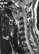
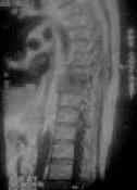
Koch's-CV junction
|
Koch's -archnoidits
|
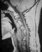
Koch's cervical
spine with cold abscess
|
|
|
|
|
|
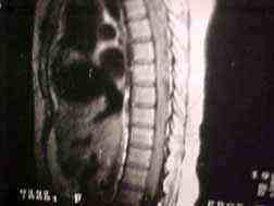
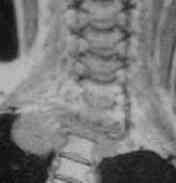
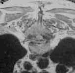
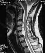
Koch's dorsal
spine
|
Koch's dorsal spine
with cold abscess
|
Koch's neural arch
(laminae involvement)
|
|
MRI :
With its high resolution, direct multiplanar imaging,
detection of early lesions and also associated lesions such as
abscesses, skip lesions and epi and intradural involvement, MRI is the
obvious choice of investigation. Contrast MRI aids in better
delineation and also in differentiating the lesion from the surrounding
edema. T1 images show decreased signal from the lesion within the 30
days and narrowing of the disc space and also loss of signal from the
nuclear pulpous. T2 may show increased signal from the involved body
and the disc narrowing with normal decreased or increased signal higher
than normally seen. Response to
|
|
|
therapy may be
seen as an increase in the signal intensity of T1 compared to previous
images.
|
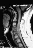
Epidural abcess-MRI T1
|
|
Treatment:
Medical:
Conservative therapy is advised by many. Bed rest and
antitubercular therapy alone have been found sufficient in most cases
including early cases of paraparesis. Bed rest is advised for 4-6 weeks
till the pain and spasm disappear and general health improves. They are
then allowed to get up, but wear braces which can be discarded after
6-8 weeks.
|
|
|
The chemotherapy
is continued for 18 months.
|
Epidural
abcess- MRI T2
|
Chemotherapy is similar to intracranial tuberculosis:
Drug:
Dosage:
Side effects:
Rifampicin:
10mgms/kg
Liver toxicity
Isoniazid: 10mgms/kg peripheral
neuritis
Pyrizanamide: 20mgms/kg Liver toxicity.
Ethambutol:
15mgms/kg.
Optic neuritis.
Surgery:
A diagnostic Ct guided needle biopsy is routine in well
established centers. Other indications are:
1. Neurological deficit which is not improving or worsening
with in 4 weeks of adequate chemotherapy - Too long a delay may lead to
problems like extradural fibrosis which may be difficult to eradicate.
2. Development of progressive neurological signs while on
adequate therapy.
3. Rapid onset paraplegia and in patients in an advanced
stage of disease when delay is risky.
4. Posterior spinal disease (because it is rare).
5. Late onset paraparesis - usually the results are less
satisfactory in healed cases. Patients with active disease respond
better.
6. Correction of kyphosis which has not responded
satisfactorily to braces and proper posturing.
Surgery may involve
1. Simple drainage of the cold abscess which would be
sufficient in these cases when the tension inside the abscess is the
cause of cord compression.
2. A direct approach thro an anterior or lateral route and
radical removal of the compressing elements such as debris, sequestrae or
granulation tissue with or without bone grafting. Medical Research
Council working party on tuberculosis of spine study showed that fusion
occurred earlier and in a higher proportion in the group with bone graft
but at 5 and 10 years there was little difference between the two. At 10
years there was a small reduction in the angle of kyphosis in the bone
grafted group and a small increase in the angle in the non grafted
series.
3. In some centers in developed countries and in modern Neuro/Orthopedic
practice, instrumentation has a significant place with good results and
early mobilization. The main problem is the formation of a focus of
infection and of course the cost involved. The current trend is to use
instruementation.
4. Laminectomy is an unsatisfactory procedure except in a
few cases when the compressing element is posterior, a condition seen in
tuberculous disease of the neural arch.
5. In the case of cranio vertebral tuberculosis, urgent
skull traction to reduce the atlanto axial subluxation is mandatory. In
some such closed reduction may not be satisfactory. They require excision
of the diseased bone granulation tissue thro a transoral route followed
by a C1 - C2 posterior Fusion either at the same sitting or at a second
stage.
B) Extradural involvement:
Majority are secondary to vertebral lesion. Occasionally we
come across a lesion without any bony lesion. It is likely they are
secondary to a small hidden focus in the adjoining vertebra. The
diagnosis is often made post operatively, as there is nothing specific in
X-Ray or in MRI. The granuloma usually encircles the dura.
Laminectomy and excision followed by a complete course
of ATT is the usual practice.
C) Intradural intramedullary involvement :
Much less frequent . With absence of any indication of
tuberculosis elsewhere the diagnosis is made with histopathology.
D) Intradural extramedullary involvement:
It is the least common and the diagnosis made with
histopathology. Only eleven cases have been reported.
E) Tuberculous archnoiditis:
It is seen in patients who have had tuberculous meningitis.
Treatment is unsatisfactory. Microsurgical techniques may provide some
relief. If it is localized, intrathecal administration of hydrocortisone
or hyaluronidase have been claimed to be effective.
PROGNOSIS:
Early diagnosis with better imagings and the 2nd line
of drugs has greatly improved the prognosis without necessitating
surgery. Recurrence may be seen if the drug therapy is irregular or
discontinued after a short time, which may be the cause for the emergence
of drug resistant cases, which are on the increase lately. A number of
these cases ultimately respond to continued therapy and to carefully
worked out combinations with or without second line of drugs.
|
