|
Thoracic disc prolapse is rare an event due to the bony
thoracic cage permitting limited movements, the anterior-posterior
direction of the apophyseal joints, and relatively small size of thoracic
disc. The first description can be traced back to 1838 by key et al and
the incidence was described ass 0.04 % of disc prolapses. Incidence of
clinically significant disc is1/year/10 lakh populations. It is found
more common in people carrying loads in the back. There is no sex
predilection. Both occupation and trauma appear to be related only
incidentally in most cases, although trauma sometimes seems to be an
important precipitating and aggravating factor.
The common site is in lower third of thoracic level, the
most common being at T11 level. Multiple level protrusions are even
rarer. Only a couple of sequestrated thoracic disc is reported.
Pathophysiology:
The thoracic canal is small with little leeway between the
thoracic disc and spinal cord.
The thoracic cord is restrained from backward displacement
by the dentate ligaments.
Circulation to the lower thoracic region is precarious and
largely due to single artery of Adamkiewicz which usually arises between
T8 and L4 on the left side in 60% of cases.
The combination of mechanical and vascular damage may
account for the severe neurological deficit and poor post operative
recovery.
Vascular damage may account for a higher clinical level that
seem inappropriate for the level of radiological lesion.
Clinical features:
The history is usually vague misleading. The common symptoms
are axial pain (77%) radicular pain (64%), signs of cord symptoms (59%)
and sensory loss (36%);myelopathy are more with central herniation.
The pain may be unilateral or bilateral and generally mild
or moderate. Patients with bilateral pain tend to progress rapidly
towards a transverse myelitis. Pain may be absent.
Subjective sensory changes in association with minimal motor
deficit are highly suggestive. Sensory symptoms may be segmental,
unilateral, or bilateral. It usually begins peripherally and ascends
gradually. Decreased pain and temperature as well as hyperesthesia and
paresthesia are common.
Bladder disturbances and impotence, trohic ulcers are late
features.
History of spontaneous remission as exacerbation of symptoms
is not as common as with lumbar and cervical disc prolapses. The majority
give a history of many years duration.
Upper thoracic disc prolapses may present with brachialgia
or horner’s syndrome.
Mild scolosis or kyphosis may be present.
|
Investigations:
Plain x-ray may show reduced disc space with bony spurs.
Prolapsed disc may be calcified and seen in plain x-ray.
CT and CT myelography have become obsolete now.
MRI has revolutionized the management of thoracic disc
prolapse. Accuracy of MRI is more than 95%, depicting the extent,
relationship to cord & root and differentiating from tumors
ossified posterior longitudinal ligament and calcified ligamentum flavum.
Absence of CSF both anterior and posterior to the cord indicates
mechanical significant compression. Thus MRI is an
|

|
|
|
excellent
screening and diagnostic investigation of choice.
|
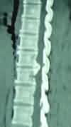
Thoracic disc--MRI
|
Thoracic
disc-CT
|
Treatment:
Asymptomatic or incidental thoracic disc does not require
treatment except for a regular follow up.
Radiological evidence of cord and / or root compression with
corresponding clinical features mandates early surgical decompression.
Routine laminectomy has been associated
with great risk.
Many approaches have been described, depending on the level
of heniation, laterality of disc prolapse, and single or multiple
levels of disc prolapses and most of all, surgeon's familiarity. The
latest addition is an endoscopic approach. The goals of
these approaches being visualization of the herniated disc without
retraction of the already deformed cord as it can not tolerate any
additional deformation.
Thoracic disc surgery is described as the most devastating
of all the disc surgeries. The preoperative diagnosis and levels
must be accurate in lateral & anterolateral approaches. Thoracic
canal being narrow, thoracic cord is very sensitive to cord compression
& very susceptible to vascular compromise. Drilling helps in decompression
without introducing instruments in the tight canal. Dissection
should be carried out under good visualization with a microscope.
Costotransversectomy
(lateral extrapleural approach):
A right sided approach is preferred to avoid artery of Adamkiewicz
unless there are lateralizing features. The patient in the partial
decubitous position with a 30-degree elevation, a long curved paraspinal
incision is made. The muscles are retracted and the rib to be
removed is identified. The intercostal neurovascular complex is separated
from its inferior. The head and neck of the rib along with a part of the
shaft is removed and the intercostal vein and arteries are followed to
the nerve root foramen. The parietal pleura is separated from the
adjacent ribs and spines. Parts of the pedicles are removed
with a drill and the dura and disc are identified and the disc is
removed. Prior to closure, the lungs are inflated. Chest tube may not be
necessary.
This approach gives a better access to the spinal canal than
the following transthoracic approach which is more popular.
Transthoracic approach:
It is the most popular approach.
A standard right posterior thoracotomy is
made.
The corresponding rib is either removed or retracted and neurovascular
bundle is followed to the intervertebral foramen.
The pleura is reflected; the pedicles are removed; and the disc is
removed as in costotransversectomy approach. The parietal pleura is
sutured over the vertebral body and chest tubes are placed. Prior to
closure, the lungs are inflated.
Central disc herniations between T-2 and T-5 may
require an anterior trans-sternal approach, the lower extent of this
approach is limited by the aortic arch.
Posterolateral approach:
A laminectomy is performed with a high speed drill. A
portion of the lateral wall of the spinal canal is drilled away, if
necessary through an horizontal skin incision at the level of the disc.
The aim is to get a lateral approach to the disc which is
removed.
Transpedicle
approach:
Through a midline incision, the paravertebral muscles are
retracted far enough to expose the facet joints. The facets and the
pedicle of the vertebra caudal to the disc are removed. The
interspace is entered, and the disc is removed. If necessary, laminectomy
is performed after disc excision.
It is a simple procedure and the results are
encouraging.
Endoscopic discectomy is being employed in
certain centers; discectomies, corpectomies, and instrumented fusions
have been performed thoracoscopically.
Surgical Results & Complications
Best results are obtained in patients with only
radicular pain with or without mild signs of myelopathy.
Severe preoperative deficit long duration of symptoms
carries a poor prgnosis.
Various reports suggest satisfactory pain relief in 79%;
improved myelopathy in 71 to 97%; improvement in sphincter functions in
60%.
|
